SI Joint Dysfunction: A Hidden Source of Lower Back, Hip, and Leg Pain

Chronic lower back or hip pain can feel overwhelming—especially when standard treatments fail to provide answers. One frequently missed cause is Sacroiliac (SI) Joint Dysfunction, a condition that often flies under the radar because its symptoms closely resemble other spinal and hip disorders.
The sacroiliac joints serve as the critical connection between the spine and pelvis. When they fail to function correctly, pain can persist for years, leading to repeated misdiagnoses, emotional distress, and reduced mobility. Understanding SI joint dysfunction—from anatomy to treatment—can dramatically improve outcomes for patients and clinicians alike.
Research suggests that 15–30% of chronic lower back pain cases may originate from the SI joints. Women are disproportionately affected, particularly during pregnancy and the postpartum period, due to hormonal changes and pelvic instability. Athletes, individuals with joint hypermobility, and those with connective tissue disorders such as Ehlers-Danlos Syndrome (EDS) also face increased risk.
Anatomy of the Sacroiliac Joints
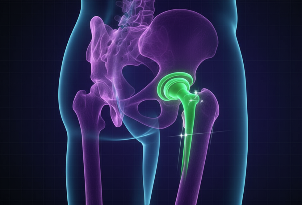
The sacroiliac joints are a pair of strong, weight-bearing joints formed where the sacrum (the triangular bone at the base of the spine) meets the ilium bones of the pelvis. Despite their small range of motion, these joints play a major role in transferring forces from the upper body to the legs.
Their stability depends on dense ligaments and surrounding muscles. While the SI joints allow only minimal movement, even slight disruptions in alignment or motion can lead to pain and dysfunction.
What Is SI Joint Dysfunction?
SI joint dysfunction occurs when the joint moves too much (hypermobility) or too little (hypomobility). This abnormal motion interferes with proper load distribution, triggering inflammation, muscle strain, and referred pain.
Importantly, SI joint dysfunction is typically a mechanical problem, not a structural defect. Imaging tests often appear normal, making clinical evaluation essential.
Common Causes and Risk Factors
Several factors can contribute to SI joint dysfunction, including:
-
Falls, accidents, or direct trauma
-
Pregnancy and childbirth
-
Uneven leg length
-
Prior spinal or pelvic surgery
-
Repetitive activities such as running or stair climbing
-
Inflammatory conditions like ankylosing spondylitis
-
Joint hypermobility disorders (e.g., EDS)
Symptoms of SI Joint Dysfunction
Pain related to SI joint dysfunction is often one-sided and centered in the lower back or buttock. It may radiate into the hip, groin, or thigh, frequently being mistaken for sciatica or disc-related pain.
Common complaints include:
-
Pain during walking, standing, or climbing stairs
-
Increased discomfort after prolonged sitting
-
Trouble sleeping on one side
-
Sharp pain when rising from a seated position
How the Body Compensates
When one SI joint malfunctions, the body adapts in ways that can worsen symptoms. These compensations may lead to:
-
Chronic lower back strain
-
Hip or knee pain
-
Overuse injuries on the opposite side
-
Tight hamstrings or hip flexors
-
Achilles tendon irritation
-
Tingling or numbness in the thigh or calf
Over time, these patterns reinforce a cycle of pain and dysfunction.
Activities That Worsen SI Joint Pain
Certain movements and habits can aggravate SI joint symptoms, including:
-
Sitting or standing for extended periods
-
Heavy lifting
-
Twisting or pivoting motions
-
High-impact exercise
-
Frequent stair climbing
Without intervention, complications may include persistent stiffness, muscular imbalance, and chronic pain syndromes.
Why SI Joint Dysfunction Is Often Missed
Diagnosing SI joint dysfunction is challenging for several reasons:
-
Symptoms overlap with spine, hip, and nerve disorders
-
There is no single definitive diagnostic test
-
Pain patterns vary widely among patients
-
Imaging studies are often inconclusive
-
Limited awareness among healthcare providers
Because of these challenges, SI joint pain is frequently attributed to herniated discs, piriformis syndrome, or sciatica.
How SI Joint Dysfunction Is Diagnosed
Medical History
A thorough history includes prior injuries, pregnancies, surgeries, and activity levels.
Physical Examination
Clinicians perform specific stress tests that provoke SI joint pain, such as:
-
FABER (Flexion, Abduction, External Rotation)
-
Patrick’s test
-
Gaenslen’s test
-
Thigh thrust test
-
Pelvic compression test
When three or more tests are positive, SI joint involvement becomes highly likely.
Imaging Studies
X-rays and MRI scans help rule out fractures, infections, or inflammatory diseases but may not directly confirm SI joint dysfunction.
Diagnostic SI Joint Injection
Image-guided injections of local anesthetic into the SI joint are considered the most reliable diagnostic tool. Significant pain relief following injection strongly supports the diagnosis.
Conditions That Mimic SI Joint Dysfunction
Several disorders can resemble SI joint pain, including:
-
Ankylosing spondylitis – inflammatory arthritis affecting the spine and SI joints
-
Sacroiliitis – true inflammation of the SI joint due to infection or autoimmune disease
-
Lumbar disc herniation – nerve compression causing radiating leg pain
-
Piriformis syndrome – sciatic nerve compression in the deep gluteal region
-
Sciatica – nerve-root irritation originating in the lumbar spine
Accurate differentiation is essential for effective treatment.
Treatment Options for SI Joint Dysfunction
Management typically progresses from conservative care to advanced interventions only if necessary.
Conservative (Non-Surgical) Treatment
Physical Therapy
Physical therapy is the foundation of treatment. Programs focus on:
-
Core stabilization
-
Pelvic alignment
-
Strengthening gluteal and abdominal muscles
-
Stretching tight hip flexors and hamstrings
Medications and Thermal Therapy
NSAIDs help control inflammation, while heat and ice therapy can reduce muscle tension and swelling.
SI Joint Belts
Pelvic belts or braces provide external stabilization, limiting excessive joint motion and easing pain during daily activities.
Manual Therapy
Chiropractic or osteopathic techniques may improve joint mechanics and muscle balance when performed by trained professionals.
Many patients achieve substantial improvement with these conservative strategies alone.
Minimally Invasive Procedures
When conservative care fails, additional options include:
Steroid Injections
Image-guided corticosteroid injections deliver targeted anti-inflammatory relief, often improving mobility and reducing pain temporarily.
Radiofrequency Ablation (RFA)
RFA uses heat to disrupt pain-transmitting nerves around the SI joint, providing relief that may last several months or longer.
Surgical SI Joint Fusion
For persistent pain lasting longer than six months despite appropriate treatment, SI joint fusion may be considered.
This minimally invasive procedure stabilizes the joint using implants or screws, eliminating painful micromovement. While many patients experience lasting relief, surgery carries risks and is reserved for carefully selected cases.
Regenerative and Biologic Therapies
Stem Cell Therapy
Stem cells harvested from bone marrow or fat tissue are injected into the SI joint to promote tissue repair and reduce inflammation.
Platelet-Rich Plasma (PRP)
PRP uses concentrated growth factors from the patient’s own blood to support healing and joint health.
Although early studies are promising, these treatments remain investigational and should be pursued through reputable medical centers.
Exercises That May Help SI Joint Pain
Under professional guidance, the following exercises may improve stability:
-
Pelvic tilts
-
Glute bridges
-
Single knee-to-chest stretch
-
Hip adduction and abduction strengthening
-
Figure-4 stretch
Exercises to Avoid
To prevent flare-ups, avoid:
-
Running or jumping
-
Deep squats
-
Uneven or asymmetrical movements without supervision
Advances in SI Joint Research
Recent studies highlight the importance of muscle imbalance in sustaining SI joint dysfunction. Early, targeted rehabilitation focusing on biomechanics yields better long-term results.
Additionally, advances in imaging and AI-assisted diagnostics may soon improve differentiation between SI joint pain and other spinal conditions.